

Call us today on 0800 724 633 or email info@scienze.nz to order and experience the AAT Bioquest Assay Advantage!

LysoBrite Image Example

Images above of HeLa cells stained with AAT’s LysoBrite ™ Green (left), and Invitrogen’s LysoTracker® Green DND-26 (right) in a Costar black wall/clear bottom 96-well plate. Samples were continuously illuminated for 120 seconds, and the signals were compared before and after the exposure by using a Keyence fluorescence microscope.
LysoBrite™ Dye Range
Lysosomes are cellular organelles which contain acid hydrolase enzymes to break up waste materials and cellular debris. Lysosomes digest excess or worn-out organelles, food particles, and engulfed viruses or bacteria. Find the following range of LysoBrite™Dyes:


LysoBrite™ Dyes Safety Data Sheet


For more information please email info@scienze.nz and one of our technical specialists will contact you directly to discuss your needs.
LysoBrite™ NIR Overview
| Catalog Number: |
22641
|
| Unit Size: |
500 Tests
|
| Molecular weight | Excitation (nm) | Emission (nm) |
| 730.89 | 634 | 649 |
Lysosomes are cellular organelles which contain acid hydrolase enzymes to break up waste materials and cellular debris. Lysosomes digest excess or worn-out organelles, food particles, and engulfed viruses or bacteria. The membrane around a lysosome allows the digestive enzymes to work at pH 4.5. The interior of the lysosomes is acidic (pH 4.5-4.8) compared to the slightly alkaline cytosol (pH 7.2). The lysosome maintains this pH differential by pumping protons from the cytosol across the membrane via proton pumps and chloride ion channels. LysoBrite™ NIR selectively accumulates in lysosomes probably via the lysosome pH gradient. The lysotropic indicator is a hydrophobic compound that easily permeates intact live cells, and trapped in lysosomes after it gets into cells. Its fluorescence is significantly enhanced upon entering lysosomes. This key feature significantly reduces its staining background and makes it useful for a variety of studies, including cell adhesion, chemotaxis, multidrug resistance, cell viability, apoptosis and cytotoxicity. It is suitable for proliferating and non-proliferating cells, and can be used for both suspension and adherent cells. LysoBrite™ dyes significantly outperform the equivalent LysoTracker ™dyes (from Invitrogen). LysoBrite™ dyes can stay in live cells for more than a week with very minimal cell toxicity while the LysoTracker dyes can only be used for a few hours.
Calculators
Common stock solution preparation
| 0.1 mg | 0.5 mg | 1 mg | 5 mg | 10 mg | |
| 1 mM | 136.819 µL | 684.097 µL | 1.368 mL | 6.841 mL | 13.682 mL |
| 5 mM | 27.364 µL | 136.819 µL | 273.639 µL | 1.368 mL | 2.736 mL |
| 10 mM | 13.682 µL | 68.41 µL | 136.819 µL | 684.097 µL | 1.368 mL |

Figure 1. Image of HeLa cells stained with Cell Navigator™ Lysosome Staining Kit in a Costar black wall/clear bottom 96-well plate.
LysoBrite™ Green Overview
| Catalog Number: |
22643
|
| Unit Size: |
500 Tests
|
| Molecular weight | Excitation (nm) | Emission (nm) |
| 453.34 | 500 | 509 |
Lysosomes are cellular organelles which contain acid hydrolase enzymes to break up waste materials and cellular debris. Lysosomes digest excess or worn-out organelles, food particles, and engulfed viruses or bacteria. The membrane around a lysosome allows the digestive enzymes to work at pH 4.5. The interior of the lysosomes is acidic (pH 4.5-4.8) compared to the slightly alkaline cytosol (pH 7.2). The lysosome maintains this pH differential by pumping protons from the cytosol across the membrane via proton pumps and chloride ion channels. LysoBrite™ Green selectively accumulates in lysosomes probably via the lysosome pH gradient. The lysotropic indicator is a hydrophobic compound that easily permeates intact live cells, and trapped in lysosomes after it gets into cells. Its fluorescence is significantly enhanced upon entering lysosomes. This key feature significantly reduces its staining background and makes it useful for a variety of studies, including cell adhesion, chemotaxis, multidrug resistance, cell viability, apoptosis and cytotoxicity. It is suitable for proliferating and non-proliferating cells, and can be used for both suspension and adherent cells.
Calculators
Common stock solution preparation
| 0.1 mg | 0.5 mg | 1 mg | 5 mg | 10 mg | |
| 1 mM | 220.585 µL | 1.103 mL | 2.206 mL | 11.029 mL | 22.058 mL |
| 5 mM | 44.117 µL | 220.585 µL | 441.17 µL | 2.206 mL | 4.412 mL |
| 10 mM | 22.058 µL | 110.292 µL | 220.585 µL | 1.103 mL | 2.206 mL |

Figure 1. Images of HeLa cells stained with A: AAT’s LysoBrite ™ Green, B: Invitrogen’s LysoTracker® Green DND-26 in a Costar black wall/clear bottom 96-well plate. Samples were continuously illuminated for 120 seconds, and the signals were compared before and after the exposure by using a Keyence fluorescence microscope.
LysoBrite™ Red Overview
| Catalog Number: |
22645
|
| Unit Size: |
500 Tests
|
| Molecular weight | Excitation (nm) | Emission (nm) |
| 698.94 | 575 | 595 |
Calculators
Common stock solution preparation
| 0.1 mg | 0.5 mg | 1 mg | 5 mg | 10 mg | |
| 1 mM | 143.074 µL | 715.369 µL | 1.431 mL | 7.154 mL | 14.307 mL |
| 5 mM | 28.615 µL | 143.074 µL | 286.148 µL | 1.431 mL | 2.861 mL |
| 10 mM | 14.307 µL | 71.537 µL | 143.074 µL | 715.369 µL | 1.431 mL |

Figure 1. Images of HeLa cells stained with A: LysoBrite ™ Red, B: LysoTracker® Red DND-99 (from Invitrogen) in a Costar black wall/clear bottom 96-well plate. Samples were continuously illuminated for 120 seconds, and the signals were compared before and after the exposure by using an Olympus fluorescence microscope.

Figure 2. Lysosome localization and motility is altered for starvation-induced Hela cells. A: Healthy untreated Hela cells. Lysosomes (Red) were dispersed widely throughout the cytosol in cells. B: Starved Hela Cells. The cells were starved for 24 hours (no serum), and lysosomes were aggregated in the perinuclear region. Nuclei were stained with Hoechst 33342.

Figure 3. Image of Hela cells stained with Cell Navigator™ Lysosome Staining Kit in a Costar black wall-clear bottom 96-well plate.
LysoBrite™ Blue Overview
| Catalog Number: |
22642
|
| Unit Size: |
500 Tests
|
| Molecular weight | Excitation (nm) | Emission (nm) |
| 331.42 | 432 | 479 |
Lysosomes are cellular organelles which contain acid hydrolase enzymes to break up waste materials and cellular debris. Lysosomes digest excess or worn-out organelles, food particles, and engulfed viruses or bacteria. The membrane around a lysosome allows the digestive enzymes to work at pH 4.5. The interior of the lysosomes is acidic (pH 4.5-4.8) compared to the slightly alkaline cytosol (pH 7.2). The lysosome maintains this pH differential by pumping protons from the cytosol across the membrane via proton pumps and chloride ion channels. LysoBrite™ Blue selectively accumulates in lysosomes probably via the lysosome pH gradient. The lysotropic indicator is a hydrophobic compound that easily permeates intact live cells, and trapped in lysosomes after it gets into cells. Its fluorescence is significantly enhanced upon entering lysosomes. This key feature significantly reduces its staining background and makes it useful for a variety of studies, including cell adhesion, chemotaxis, multidrug resistance, cell viability, apoptosis and cytotoxicity. It is suitable for proliferating and non-proliferating cells, and can be used for both suspension and adherent cells.
Calculators
Common stock solution preparation
| 0.1 mg | 0.5 mg | 1 mg | 5 mg | 10 mg | |
| 1 mM | 301.732 µL | 1.509 mL | 3.017 mL | 15.087 mL | 30.173 mL |
| 5 mM | 60.346 µL | 301.732 µL | 603.464 µL | 3.017 mL | 6.035 mL |
| 10 mM | 30.173 µL | 150.866 µL | 301.732 µL | 1.509 mL | 3.017 mL |

Figure 1. Image of HeLa cells stained with Cell Navigator™ Lysosomal Staining Kit in a Costar black wall/clear bottom 96-well plate.
LysoBrite™ Orange Overview
| Catalog Number: |
22644
|
| Unit Size: |
500 Tests
|
| Molecular weight | Excitation (nm) | Emission (nm) |
| 670.89 | 542 | 564 |
Lysosomes are cellular organelles which contain acid hydrolase enzymes to break up waste materials and cellular debris. Lysosomes digest excess or worn-out organelles, food particles, and engulfed viruses or bacteria. The membrane around a lysosome allows the digestive enzymes to work at pH 4.5. The interior of the lysosomes is acidic (pH 4.5-4.8) compared to the slightly alkaline cytosol (pH 7.2). The lysosome maintains this pH differential by pumping protons from the cytosol across the membrane via proton pumps and chloride ion channels. LysoBrite™ Orange selectively accumulates in lysosomes probably via the lysosome pH gradient. The lysotropic indicator is a hydrophobic compound that easily permeates intact live cells, and trapped in lysosomes after it gets into cells. Its fluorescence is significantly enhanced upon entering lysosomes. This key feature significantly reduces its staining background and makes it useful for a variety of studies, including cell adhesion, chemotaxis, multidrug resistance, cell viability, apoptosis and cytotoxicity. It is suitable for proliferating and non-proliferating cells, and can be used for both suspension and adherent cells. LysoBrite™ dyes significantly outperform the equivalent LysoTracker ™dyes (from Invitrogen). LysoBrite™ dyes can stay in live cells for more than a week with very minimal cell toxicity while the LysoTracker dyes can only be used for a few hours. LysoBrite™ dyes are much more photostable than the LysoTracker dyes.
Calculators
Common stock solution preparation
| 0.1 mg | 0.5 mg | 1 mg | 5 mg | 10 mg | |
| 1 mM | 149.056 µL | 745.279 µL | 1.491 mL | 7.453 mL | 14.906 mL |
| 5 mM | 29.811 µL | 149.056 µL | 298.111 µL | 1.491 mL | 2.981 mL |
| 10 mM | 14.906 µL | 74.528 µL | 149.056 µL | 745.279 µL | 1.491 mL |
Fi
Figure 1. HeLa cells were incubated in 1X HBSS buffer with 5% serum to induce starvation. Following starvation, cells were treated with Autophagy Green™ (Cat No. 23002) working solution for 20 minutes in a 37°C, 5% CO2 incubator and then washed 3 times. Nuclei were labeled with Hoechst 33342 (Cat No. 17530). Lysosomes were labeled with LysoBrite™ Orange (Cat No. 22657).

Figure 2. Image of HeLa cells stained with Cell Navigator™ Lysosomal Staining Kit in a Costar black wall/clear bottom 96-well plate using an Olympus fluorescence microscope TRITC channel.
LysoBrite™ Deep Red Overview
| Catalog Number: |
22646
|
| Unit Size: |
500 Tests
|
| Molecular weight | Excitation (nm) | Emission (nm) |
| 746.99 | 595 | 618 |
Lysosomes are cellular organelles which contain acid hydrolase enzymes to break up waste materials and cellular debris. Lysosomes digest excess or worn-out organelles, food particles, and engulfed viruses or bacteria. The membrane around a lysosome allows the digestive enzymes to work at pH 4.5. The interior of the lysosomes is acidic (pH 4.5-4.8) compared to the slightly alkaline cytosol (pH 7.2). The lysosome maintains this pH differential by pumping protons from the cytosol across the membrane via proton pumps and chloride ion channels. LysoBrite™ Deep Red selectively accumulates in lysosomes probably via the lysosome pH gradient. The lysotropic indicator is a hydrophobic compound that easily permeates intact live cells, and trapped in lysosomes after it gets into cells. Its fluorescence is significantly enhanced upon entering lysosomes. This key feature significantly reduces its staining background and makes it useful for a variety of studies, including cell adhesion, chemotaxis, multidrug resistance, cell viability, apoptosis and cytotoxicity. It is suitable for proliferating and non-proliferating cells, and can be used for both suspension and adherent cells. LysoBrite™ dyes significantly outperform the equivalent LysoTracker ™dyes (from Invitrogen). LysoBrite™ dyes can stay in live cells for more than a week with very minimal cell toxicity while the LysoTracker dyes can only be used for a few hours. LysoBrite™ dyes can survive a few generations of cell division. In addition, LysoBrite™ dyes are much more photostable than the LysoTracker dyes.
Calculators
Common stock solution preparation
| 0.1 mg | 0.5 mg | 1 mg | 5 mg | 10 mg | |
| 1 mM | 133.871 µL | 669.353 µL | 1.339 mL | 6.694 mL | 13.387 mL |
| 5 mM | 26.774 µL | 133.871 µL | 267.741 µL | 1.339 mL | 2.677 mL |
| 10 mM | 13.387 µL | 66.935 µL | 133.871 µL | 669.353 µL | 1.339 mL |
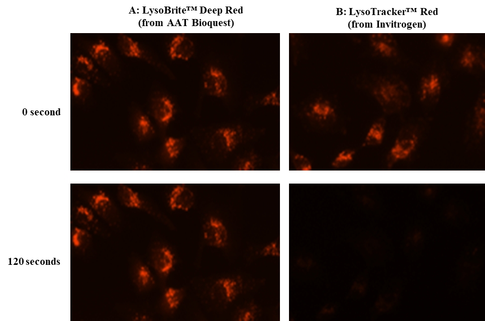
Figure 1. Image of Hela cells stained with the A: LysoBrite™ Deep Redor B: LysoTracker® Red DND-99 (from Invitrogen) in a Costar black 96-well plate. The TRTIC signals were compared at 0 and 120 seconds exposure time by using an Olympus fluorescence microscope.

Figure 2. Image of Hela cells stained with LysoBrite™ Deep Red.
LysoBrite™ Red DND-99 Overview
| Catalog Number: |
22647
|
| Unit Size: |
500 Tests
|
| Molecular weight | Excitation (nm) | Emission (nm) |
| 399.25 | 573 | 592 |
LysoBrite Red DND-99 is chemically same to the LysoTracker® Red DND-99 used for labeling and tracking acidic organelles in live cells (LysoTracker® is the trademark of ThermoFisher). It has good selectivity for acidic organelles, and reasonably retaining its staining pattern after aldehyde fixation. The LysoBrite™ probes consist of a fluorophore linked to a weak base that is only partially protonated at neutral pH, allowing them to freely permeate cell membranes to label live cells.
Calculators
Common stock solution preparation
| 0.1 mg | 0.5 mg | 1 mg | 5 mg | 10 mg | |
| 1 mM | 250.47 µL | 1.252 mL | 2.505 mL | 12.523 mL | 25.047 mL |
| 5 mM | 50.094 µL | 250.47 µL | 500.939 µL | 2.505 mL | 5.009 mL |
| 10 mM | 25.047 µL | 125.235 µL | 250.47 µL | 1.252 mL | 2.505 mL |

Figure 1. Chemical structure for LysoBrite™ Red DND-99.
For more information please email info@scienze.nz and one of our technical specialists will contact you directly to discuss your needs.
Excellence in Scientific Solutions
A to Z Product Listing
A to Z Product Listing
Copyright © 2025, Scienze. All rights reserved.